A Medical platform specially related to Radiology field and also knowledgeable site, for basic to depth Medical world

What is Kilogram?

Kilogram (kg)


-The kilogram is unit of mass; it is equal to the mass of the international prototype of the kilogram.
-It is equal to the mass of the international prototype of the kilogram.
-The kilogram is the base unit of mass in the international system of units.
What is Kilogram?
Fig.1 Kilogram


Unit of:    mass. 

Unit system:    SI base unit.

Symbol- kg
Share:

What is Meter, Definition of Meter, Meter Symbol | Radiology Buzz

Meter, Definition of Meter


The meter is international system (SI) of units.
-  The meter or meter is the base unit of length in some metric system.
-  Meter SI unit symbol is m.
-  A piece of equipment that measures the amount of gas, water, electricity, voltage, etc.
Meter
Fig.1 Meter


1 meter in....                                  Is equal to.....


SI units                                                  1000 mm

                                                               0.001 km

imperial/US Units                                 1.0936 yd

                                                                              3.2808 ft
                                                                              39.370 in

Nautical units                                0.000539996 nmi

Unit system:                                  SI base unit.

Unit of:                                             length

Symbol:                                            m

Share:

What is X-Ray Properties, Properties of X-Ray Radiation, Properties of X-Ray Photons, Uses of X-Ray | Radiology Buzz

X-Rays Properties


1. X-rays are very energetic form of electromagnetic radiation.

2. They passes great penetrating power. X-ray penetrate different object more or less according to their density.

3. X-rays are used to view images of the bones and other structure in the body.

4. X-rays are not influenced by electric or magnetic field.

5. However they can cause ionization of gases indirectly as they can remove orbital electron from atoms.

6. Their wavelength ranges from 0.1 to 0.5 Angstrom (0.01 to 10 nanometer ), corresponding to frequencies in the range 30 petahartz to 30 exahertz ( 3 × 10¹⁶ Hz to 3 × 10¹⁹ Hz ) and Energies in the range 100 ev to 100 kev.

7. They travel at the speed of light i.e. ( 3 × 10 m/s ).

8. They travel in straight line, like light rays, however, unlike light rays, they cannot be focused by a lens.

9. They liberate minute amounts of heat on passing through matter.

10. They cause fluorescence of certain crystals.

11.They cause photographic effect on silver halide crystals, which is a chemical change.

12. They can produce other chemical as well as biological changes mainly by ionization and excitation.

What is X-Ray Properties?
Fig.1:  X-Ray Properties
13. Being electromagnetic waves, they are electrically neutral.

14. X-rays affect the photographic film and form latent image which is a chemical changes.

15. X-rays are invisible radiation.

16. X-rays produce chemical changes in substances through which the pass.

17. X-rays produce biological effects in living organism.

18. The cells can be either damaged or killed due to x-ray exposure.

19. X-rays produce fluorescence in material like calcium tungstate and zinc calcium sulfide etc.
Share:

Production of X-Rays, Production of an x ray beam, x ray production focusing of electrons | Radiology Buzz

PRODUCTION OF X-RAYS


X-ray are produced by energy conversion when a fast moving stream of electron is suddenly decelerated in the Target Anode of an x-ray tube.
Production of X-Ray
Fig.1 X-Ray Tube

The x-ray tube is made of Pyrex glass that enclose a vacuum containing two electrodes (This is a diode tube).

Diode Definition - An electronic device in which the electric current flows in one direction only.

Electrons are produced by the heated tungsten filament and accelerated across the tube to hit the tungsten target, where x-ray are produced.

This section will describe the design of the x-ray tube and will review the way in which x-rays are produced.

The electrodes are designed so that electrons produced at the Cathode (negative electrode of filament) can be accelerated by a high potential difference to ward the Anode (Positive or Target electrode).


POTENTIAL DIFFERENCE - The difference of SUMMARY-


1. A current is passed through the tungsten filament and heat it up.


2.As it is heated up the increased energy enables electrons to be released from the filament trough Thermionic emission.

THERMIONIC EMISSION- The process by which free electrons are emitted from the surface of a metal when external heat energy is applied is called thermionic emission.

3-The electrons are attracted towards the positively charged anode and hit the tungsten target with a maximum energy determined by the tube potential (Voltage).

4-As the electrons bombard the target they interact via bremsstrahlung and characteristic interaction which result in conversion of energy into heat (99%) and x-ray photons (1%).

5-The x-ray photons are released in a beam with a range of energies (x-ray spectrum) out of the window of the tube and form the basis for x-ray image formation.


Share:

History of X-Rays, The First X-Ray, X-Rays Discovery, X-Ray History Timeline | Radiology Buzz

HISTORY OF X-RAYS

X-Ray were discovered in 8 November 1895 by Sir Wilhelm Conrad  Roentgen (1845-1923)who was professor at Wuerzburg University in Germany.

History of X-Rays
Sir Wilhelm Conrad Roentgen (1845-1923)
Roentgen working with cathode ray tube in his laboratory, Roentgen observed crystal near a high voltage cathode ray tube exhibiting a fluorescent glow, ever when he shielded them with dark paper. some form of energy was being produced by the tube and it was penetrating the paper and causing the crystals to glow.

Roentgen placed various objects between the tube and the screen, and the screen still glowed. finally, he put his hand in front of the tube, and saw the outline of his bones projected into the fluorescent screen. Immediately after discovering x-rays themselves, he had discovered their most beneficial application.

History of X-Rays
Roentgen's wife Bertha's Hand
The first x-ray photograph Roentgen’s wife Bertha’s hand.

Roentgen first publication about his discovery was released in the days that followed on 28 December 1895 it was entitled “On a new kind of rays”.

He suggested the name “X-ray” for his discovery, arguing that since the origin of the rays was unknown, it was appropriate to refer to them as X, the mathematical term for the unknown.
For the discovery, Roentgen was awarded the first noble prize in physics, in 1901 (During world war) x-ray were already being used for medical purposes.
Share:

PET scan: Definition purpose, side effect, procedure, Advantage, Disadvantage | Radiology Buzz


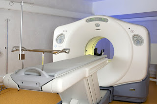
(PET SCAN) POSITRON EMISSION TOMOGRAPHY

Introduction 

An imaging modality which gives better functional and morphological information in combination with CT and MRI.

Used radioactive traces such FDG (Fluorodeoxyglucose)as  it is either injected into the blood stream, inhaled or swallowed.

What is PET Scan?
Fig.1   PET SCAN MACHINE

It is a non invasive technique.

Past of nuclear medicine scan.

Provides metabolic information of neoplastic processes but is not specific for neoplastic processes.

It accumulation in physiologically active areas in various organs including brain, muscles, salivary glands, myocardium, GIT, brown adipose tissue, thyroid gland & gonads.

Why PET Scan are used?

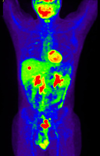
The glucose in FDG is taken up by these active tissue for metabolism which in turn carries the radioactive traces tagged with the glucose.

Advantage of Positron Emission Tomography

Ability to study body function through biochemical functions. i.e., it is able to detect disease before changes in the anatomy becomes apparent making it more effective in diagnosing then other imaging test.
What is PET Scan?
Fig.2   PET Scan

Can be used as an alternative to biopsy and other exploratory surgeries to determine how much a disease has spread.

Its ability to distinguish between benign (non-cancerous) and malignant tool that can reduce the number of unnecessary surgeries performed due to  diagnosis and staging data.

Effective used to diagnose early stage of neurological illness such as epilepsy, Alzheimer's disease and other dementia.

Disadvantage of Positron Emission Tomography

Does not provide anatomical details. 

Large number of false positive and false negative results.

Difficult to differentiate the lesions from areas of normal physiological uptake.

Radioactive substance used may not be suitable for patients who are pregnant, suspect they are pregnant or are breast feeding.

The radioactive exposure in PET imaging means that there is only a limited amount of times a patient can undergo this procedure.

PET scan requires cyclotrons, an expensive machine that creates the radioisotopes that are used in the radioactive traces required for PET imaging.

Allergic reaction to traces.

Share:

Followers

Most Popular

Recent Posts

Pages

Contact Us

Name

Email *

Message *