A Medical platform specially related to Radiology field and also knowledgeable site, for basic to depth Medical world

Showing posts with label Physics of X-Ray. Show all posts

The Penetrating Power of Radiation

Definition of Penetrate :


  1. To go through or into something, especially when this is difficult.
  2. To discover the inner contents or meaning of.
  3. The penetrating power of radiations is the measurement of energy of those radiations. As the energy of radiations increases, the penetration power of radiations also increases.


Penetrating power of Rays:

The penetrating power of different types of radiation: 

  1. X-Ray
  2. Alpha
  3. Beta
  4. Gamma rays

X-Ray:

The penetrating power of radiation
Fig.2  Penetrating power of X-Ray
  • X-ray are electromagnetic radiation.
  • X-ray are photons or high energy light waves which are usually emitted by radium or cobalt. These are can easily penetrate our body therefore also called penetrating radiation.
  • X-ray can be stopped by a thick lead barrier and a wall.
  • Human bones or organs are deferentially penetrable.

Gamma Rays:

  • Gamma radiation are penetrating electromagnetic radiation, like X-Ray.
  • Gamma rays are emitted by the nucleus of radioactive atoms.
  • They can be used to kill cancer cell and it also used sterilise medical (Surgery) equipment.
  • Gamma rays are the high energetic radiation, with a short wavelength less than one tenth of a nano meter.
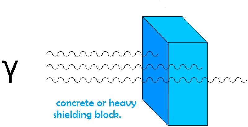
  • Gamma rays are the more difficult  to stop these rays require lead, concrete or heavy shielding block.

    GAMMA RAYS SYMBOL - 𝛄
The penetrating power of gamma rays
Fig.3 Penetrating power of gamma rays 

Alpha Rays: 


  • Alpha particles, also called alpha ray or alpha radiation. 
  • Alpha rays are the positively charge particles rays, that means this rays atoms only contains proton and no electrons.  
  • These are fast moving helium atoms.
  • It consist of two protons and two neutrons bound together into a particle identical to a helium-4 nucleus.
  • They have high energy, typically in the MeV range, but due to their large mass, they are stopped by just a few inches of air, or a piece of paper.
  • Alpha radiation is not dangerous because it is unlikely to reach living cells inside the body. Beta and gamma radiation are the most dangerous sources because they can penetrate the body tissue (skin) and damage the cells inside
    ALPHA RAYS SYMBOL - α
    penetrating power of differential of radiation
    Fig.4  Penetrating power of alpha particle


Beta Rays: 

  • These are fast moving and high energy electron.
  • It is emitted by the radioactive decay of atomic nucleus during the process of beta decay.
  • It is two forms of beta decay 𝛃¯  decay and 𝛃+ decay, which produce electrons positrons.
  • Energy 0.5 MeV
  • Beta particles are a type of ionizing radiation.


    BETA RAYS SYMBOL - 𝛃
the penetrating power of radiation
Fig.5 Penetrating Power of Beta Rays  


Share:

What is Anode?

Component of X-Ray Tube:
(ANODE)
  • The positive terminal in x-ray tube is called the anode.
  • Anode of x-ray tubes are of two types.
        1. Stationary Anode 
        2. Rotating Anode

Stationary Anode: 

  • The anode of a stationary anode x-ray tube consist of a small plate of a tungsten, 2 or 3 mm thick, that is embedded in a large mass of copper.
    What is Anode?
    Fig.1   Stationary Anode X-Ray Tube 
  • The tungsten plate is square or rectangular in shape with each dimension usually being greater than 1 cm.
  • The anode angle is usually 15⁰ to 20⁰.

Tungsten: Tungsten is chosen as the target material for several reasons.

-It has a high atomic number (74). Which makes a more efficient for the production of x-ray.- In addition, because of its, high melting point, it is able to withstand the high temperature produced.- Mostly metals melt between 300 and 1500⁰c, whereas tungsten melts at 3370⁰c.- Tungsten is a reasonably good material for the absorption of heat and for the rapid dissipation of the heat away from the target area.-The rather small tungsten target must be bonded to the much larger copper portion of the anode to facilitate heat dissipation in spite of its good thermal characteristics, tungsten cannot withstand the heat of repeated exposures.
What is Anode?
Fig.2    Stationary Anode Tube

-The size of the tungsten target is larger then the area bombarded by the electron stream. ---This is necessary because of the low melting point of copper (1070⁰c). A single x-ray exposure may raise the temperature of the bombarded area of the tungsten target by 1000⁰c or more.
-All the metals expand when heated, but they expand at different rates.


Rotating Anode:

  • The anode of a rotating anode tube consists of a large disc of tungsten, which theoretically rotates at a speed of about 3600 revolution per minute (rpm) when an exposure in being made.
  • In practice, the anode never reaches a speed of 3600 rpm because of mechanical factors such as slipping between the rotor and bearing. Therefore, to calculate the ability of a tube to withstand high loads, a peed of 3000 rpm is usually assumed.
    What is Anode?
    Fig.3 Rotating Anode X-Ray Tube
  • Functioning rotating anode will never drop below  3000 rpm, and will usually be greater than 3000 rpm, if 60 hertz (HZ) current is used.
  • The tungsten disc has a beveled edge.
  • The angle of the bevel may very from 6 to 20⁰. The bevel is used to take advantage of the line focus principle.
  • The purpose of the rotating anode is to spread the heat produced during an exposure over a large area of the anode.
  • The filament and focusing cup of the x-ray tube produce an electron beam that covers an area of the anode 7-mm high and 2-mm wide, the area of the anode bombarded by electrons is represented by a 14-mm rectangle. We recognise that a 2-mm focal spot is a useful way to illustrate a point.
  • If the bevel of the target is 16.5⁰.
  • If the anode were stationary, the entire heat load would be delivered to this one small 14-mm area of the target.
  • If the target is made to rotate at a speed 3600 rpm. however, the electron will bombard a constantly changing area of the target.
  •  To make the anode rotate, some mechanical problems must be overcome, be cause the anode is contained within the vacuum of the tube. The power to effect rotating is provided by a magnetic field produced by stator coil that surround the neck of the x-ray tube outside the envelope.
  • The magnetic field produced by the stator coils induces a current in the copper rotor of the induction motor, and this induced current provides the power to rotate the anode assembly
  • the development of rotating anode x-ray tubes, the life of the tube was quite short because of the lack of durable bearing on which the anode assembly could rotate. because of the friction produced it was necessary to lubricate the bearing, but commonly available lubricate the bearing, but commonly available lubricants could not be used.
  • Lubricants such as oil vaporise when heated and destroy the vacuum in the tube, dry lubricants such as a graphite would wear as a powder and destroy the vacuum. The problem was solved by the use of metallic lubricants (especially silver). Which are suitable for use in a high vacuum.
  • In modern rotating anode tubes, bearing wear has become a negligible factor in overall tube life.
  • Heat dissipation in a rotating anode tube presents an additional problem.
  • Heat generated in a solid tungsten disc is dissipated by radiating through the vacuum to the wall of the tube, and then into the surrounding oil and tube housing.
  • The rotating anode tube, absorption of heat by the anode assembly is undesirable because heat absorbed by the bearing of the anode assembly would cause them to expand and bind. Because of this problem the stem, which connects the tungsten target to the remainder of the anode assembly, is made of molybdenum.
  • Molybdenum has a high melting point (2600⁰) and is a poor heat conductor.
  • Thus, the molybdenum stem provides a partial heat barrier between the tungsten dic and the bearing of the anode assembly. 
Share:

What is Cathode?


COMPONENT OF X-RAY TUBE (Cathode)


  • The negative terminal of the x-ray tube is called the cathode.
  • It has got a filament (which is the source of electron for the x-ray tube).
  • The cathode has two other elements. These are the connecting wires, which supply both the voltage (Average about 10 volts and Ampere average about 3-5 Ampere) that heat the filament.
  • And a metallic Focusing cup.
  • The number (quantity) of x-rays produced depends entirely on the number of electrons that flow from the filament to the target (Anode) of the tube.
  • The x-ray tube current measured in Milliamperes (1 mA=0.001 A), refers to the number of electron flowing per second from the filament to the target.
  • The filament is made of tungsten wire, about 0.2 mm in diameter, that is coiled to form a vertical spiral about 0.2 cm in diameter and 1 cm or less in length.
  • When current flows through this fine tungsten wire, it becomes heated. when a metal is heated its atoms absorb thermal energy and some of the electrons in the metal acquire enough energy to allow them to move a small distance from the surface of the metal (normally, electron can move with in a metal, but cannot escape from it). Their escape is referred to as the process of thermionic emission.
  • The electron cloud surrounding the filament produced by thermionic emission, has been termed the “Edison effect.”
  • A pure tungsten filament must be heated to a temperature of at least (2200 degree c) to emit a useful number of electrons (Therm-ions)
  • Tungsten is not as efficient an emitting material as other material (such as alloys of tungsten) used is some electron tubes.
  • It is chosen for use is x-ray tubes, however, because it can be drown into a thin wire that is quite strong, has a high melting point (3370 degree c).
  • It has little tendency to vaporise; thus, such a filament has a reasonably long life expectancy.
  • Electron emitted from the tungsten filament form a small cloud in the immediate vicinity of the filament. This collection what is called the space charge. However in this case electrons does not move until they acquired sufficient thermal energy to overcome the force caused by space charge. Thus emission of further electrons from filament is limited by the space charge and this is called a space charge effect.
  • When electrons leave the filament the loss of negative charges causes the filament to acquire a positive charge. The filament then attracts some emitted electrons back to itself. When a filament is heated to its emission temperature, a state of equilibrium is quickly reached.
  • In equilibrium the number of electrons returning to the filament is equal to the number of electrons being emitted.
  • As a result the number of electrons in the space charge remains constant, with the actual number depending on filament temperature.
  • The high currents that can be produced by the use of thermionic emission are possible because large numbers of electrons can be accelerated from the cathode (Negative electrode) of the x-ray tube.
  • The number of electrons involved is enormous.
  • The unit of electric current is the ampere, which may be defined as the rate of “Flow” when 1 coulomb of electricity flows through a conductor in 1 sec.
  • The coulomb is the equivalent of the amount of electric charge carried by 6.25x10¹⁸ electrons. Therefore, an x-ray tube current of 100 mA (0.01 A) may be considered as the “Flow” of 6.25x10¹⁷ electrons from the cathode to the anode in 1 sec.
  • Electrons current across an x-ray tube is in one direction only (Always cathode to anode).
  • FOCUSING CUP- Which surrounds the filament. When the x-ray tube is conducting, the focusing cup is maintained at the same negative potential as the filament.
  • The focusing cup usually made of nickel.
  • Modern x-ray tubes may be supplied with a single or more commonly, a double filament, each filament consists of a spiral of wire, and mounted side by side or one above the other, with one being longer than the other.
  • It is important to understand that only one filament is used for any given x-ray exposure; the larger filament is generally used for larger exposure.
  • FOCAL SPOT- The radiation is produced in very small area on the surface of the anode known as the focal spot.
  • What is Cathode?
    A double filament contained in a focusing cup
    • Two additional filament arrangements may be seen in highly specialised x-ray tubes. A tube with three filaments (triple focus) is available. Another application is a Stereoscopic Angiographic tube, in this tube focal spot widely separated (about a 4-cm separation). When two films are exposed, using a different focal spot for each film.
    • This tube is useful in angiography when rapid exposure of multiple stereoscopic film pairs is desired. Interval as short as 0.1 sec between exposure can be obtained with stereoscopic tubes.
    • Vaporization of the filament when it is heated acts to shorten the life of an x-ray tube, because the filament will break if it becomes too thin.
    • The filament should never be heated for longer periods than necessary.
    • The focal spot is the area of the tungsten target (Anode) that is bombarded by electrons from the cathode,(Most of the energy of the electrons is converted into heat, with less than 1% being converted into x-ray).
    • LINE FOCUS PRINCIPLE- The problems posed by the need for a large focal spot to allow greater heat loading, and the conflicting need for a small focal area to produce good radiography details, were resolved in 1918 with the development of the line focus principle.
    • The size and shape of focal spot are determined by the size and shape of the electron stream when it hits the anode. The size and shape of the filament tungsten wire coil, the construction of the focusing cup, and the position of the filament in the focusing cup.
    What is Cathode?
    Tungsten
  • REGION FOR CHOOSING TUNGSTEN AS A FILAMENT -It is capable of stable electrons emission at high temperature. -It has high melting point. -It has mechanical strength, long life, and can be easily drown out into a fine filament. -It has high atomic number 74. -it has high thermal conductivity metal.
Share:

Glass Enclosure

X-Ray Tube Components

This section will describe the design of the x-ray tube and which x-ray component are used:

X-Ray tube
Fig.1     X-Ray tube 
1. Glass Enclosure (Envelope) 2. Cathode 3. Anode 4. focusing cup 5. Insulating oil 6. Window 7. Filter


1. GLASS ENCLOSURE (ENVELOPE):

  • It is necessary to seal the two electrodes of the x-ray tube in a vacuum. If gas were present inside the tube, the electrons that were being accelerated toward the anode (Target) would collide with the gas molecules, lose energy, and cause secondary electrons to be ejected from the gas molecules.
  • This production of secondary electrons could not be satisfactory controlled. their presence would result reduced speed of the electrons impinging on the target.
  • The principle was used in the design of the early so called “Gas” x-ray tubes. which contained small amounts of gas to secure as a source of secondary electrons.
  • The purpose of vacuum in the modern x-ray tubes is to allow the number and speed of the electrons to be controlled independently.
  • The shape and size of these x-ray tubes are specially designed to prevent electric discharge between the electrons.
  • The connectivity wires must be sealed into the glass wall of the x-ray tube.
  • Pyrex glass are generally used in the x-ray tube.
Share:

What is X-Ray Properties, Properties of X-Ray Radiation, Properties of X-Ray Photons, Uses of X-Ray | Radiology Buzz

X-Rays Properties


1. X-rays are very energetic form of electromagnetic radiation.

2. They passes great penetrating power. X-ray penetrate different object more or less according to their density.

3. X-rays are used to view images of the bones and other structure in the body.

4. X-rays are not influenced by electric or magnetic field.

5. However they can cause ionization of gases indirectly as they can remove orbital electron from atoms.

6. Their wavelength ranges from 0.1 to 0.5 Angstrom (0.01 to 10 nanometer ), corresponding to frequencies in the range 30 petahartz to 30 exahertz ( 3 × 10¹⁶ Hz to 3 × 10¹⁹ Hz ) and Energies in the range 100 ev to 100 kev.

7. They travel at the speed of light i.e. ( 3 × 10 m/s ).

8. They travel in straight line, like light rays, however, unlike light rays, they cannot be focused by a lens.

9. They liberate minute amounts of heat on passing through matter.

10. They cause fluorescence of certain crystals.

11.They cause photographic effect on silver halide crystals, which is a chemical change.

12. They can produce other chemical as well as biological changes mainly by ionization and excitation.

What is X-Ray Properties?
Fig.1:  X-Ray Properties
13. Being electromagnetic waves, they are electrically neutral.

14. X-rays affect the photographic film and form latent image which is a chemical changes.

15. X-rays are invisible radiation.

16. X-rays produce chemical changes in substances through which the pass.

17. X-rays produce biological effects in living organism.

18. The cells can be either damaged or killed due to x-ray exposure.

19. X-rays produce fluorescence in material like calcium tungstate and zinc calcium sulfide etc.
Share:

Production of X-Rays, Production of an x ray beam, x ray production focusing of electrons | Radiology Buzz

PRODUCTION OF X-RAYS


X-ray are produced by energy conversion when a fast moving stream of electron is suddenly decelerated in the Target Anode of an x-ray tube.
Production of X-Ray
Fig.1 X-Ray Tube

The x-ray tube is made of Pyrex glass that enclose a vacuum containing two electrodes (This is a diode tube).

Diode Definition - An electronic device in which the electric current flows in one direction only.

Electrons are produced by the heated tungsten filament and accelerated across the tube to hit the tungsten target, where x-ray are produced.

This section will describe the design of the x-ray tube and will review the way in which x-rays are produced.

The electrodes are designed so that electrons produced at the Cathode (negative electrode of filament) can be accelerated by a high potential difference to ward the Anode (Positive or Target electrode).


POTENTIAL DIFFERENCE - The difference of SUMMARY-


1. A current is passed through the tungsten filament and heat it up.


2.As it is heated up the increased energy enables electrons to be released from the filament trough Thermionic emission.

THERMIONIC EMISSION- The process by which free electrons are emitted from the surface of a metal when external heat energy is applied is called thermionic emission.

3-The electrons are attracted towards the positively charged anode and hit the tungsten target with a maximum energy determined by the tube potential (Voltage).

4-As the electrons bombard the target they interact via bremsstrahlung and characteristic interaction which result in conversion of energy into heat (99%) and x-ray photons (1%).

5-The x-ray photons are released in a beam with a range of energies (x-ray spectrum) out of the window of the tube and form the basis for x-ray image formation.


Share:

History of X-Rays, The First X-Ray, X-Rays Discovery, X-Ray History Timeline | Radiology Buzz

HISTORY OF X-RAYS

X-Ray were discovered in 8 November 1895 by Sir Wilhelm Conrad  Roentgen (1845-1923)who was professor at Wuerzburg University in Germany.

History of X-Rays
Sir Wilhelm Conrad Roentgen (1845-1923)
Roentgen working with cathode ray tube in his laboratory, Roentgen observed crystal near a high voltage cathode ray tube exhibiting a fluorescent glow, ever when he shielded them with dark paper. some form of energy was being produced by the tube and it was penetrating the paper and causing the crystals to glow.

Roentgen placed various objects between the tube and the screen, and the screen still glowed. finally, he put his hand in front of the tube, and saw the outline of his bones projected into the fluorescent screen. Immediately after discovering x-rays themselves, he had discovered their most beneficial application.

History of X-Rays
Roentgen's wife Bertha's Hand
The first x-ray photograph Roentgen’s wife Bertha’s hand.

Roentgen first publication about his discovery was released in the days that followed on 28 December 1895 it was entitled “On a new kind of rays”.

He suggested the name “X-ray” for his discovery, arguing that since the origin of the rays was unknown, it was appropriate to refer to them as X, the mathematical term for the unknown.
For the discovery, Roentgen was awarded the first noble prize in physics, in 1901 (During world war) x-ray were already being used for medical purposes.
Share:

Followers

Most Popular

Labels

Recent Posts

Pages

Contact Us

Name

Email *

Message *